Current Diagnostic Options Have Limitations
Current Diagnostic Options Have Limitations
Conventional imaging does not reliably diagnose ccRCC1-3
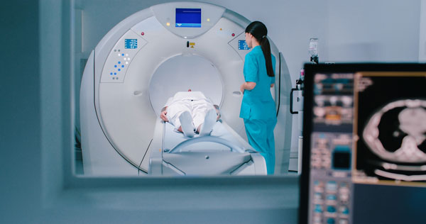
While CT and MRI are mainstays of renal imaging, neither can reliably characterize tumors as clear cell renal cell carcinoma (ccRCC). A more accurate, precise imaging approach is needed in order to optimize treatment planning.
Performance in Studies Diagnosing ccRCC4,5

More than 20% of detected masses cannot be adequately classified as benign or malignant, highlighting the need for more accurate renal imaging.6
Utility of renal mass biopsy is limited7
Although renal mass biopsy can be useful in some instances, there are several diagnostic limitations that affect its accuracy and utility in clinical practice.
Limitation
Reason

Not all patients benefit from surgery10
Although surgery is the standard of care for renal masses, there are situations where it may not be optimal.10
- Approximately one-third of resected renal masses are benign11
- ≈6,000 benign renal masses are removed annually in the United States10
References: 1. Long Z, Sun C, Tang M, et al. Single-cell multiomics analysis reveals regulatory programs in clear cell renal cell carcinoma. Cell Discov. 2022;8(1):68. 2. van Oostenbrugge TJ, Fütterer JJ, Mulders PFA. Diagnostic imaging for solid renal tumors: a pictorial review. Kidney Cancer. 2018;2(2):79-93. 3. Roussel E, Capitanio U, Kutikov A, et al. Novel imaging methods for renal mass characterization: a collaborative review. Eur Urol. 2022;81(5):476-488. 4. Lemieux S, Shen L, Liang T, et al. External validation of a five-tiered CT algorithm for the diagnosis of clear cell renal cell carcinoma: a retrospective five-reader study. AJR Am J Roentgenol. 2023;221(3):334-343. 5. Yang Z, Li M, Guo A, Liang Y, Fang P. Imaging features and clinic value of MRI and CT in diagnosis of clear cell renal cell carcinoma. Food Sci Technol (Campinas). 2022;42:e40520. 6. Butaney M, Wilder S, Patel AK, et al. Initial management of indeterminate renal lesions in a statewide collaborative: a MUSIC-KIDNEY analysis. J Urol. 2023;210(1):79-87. 7. de Silva S, Lockhart KR, Aslan P, et al. Differentiation of renal masses with multi-parametric MRI: the de Silva St George classification scheme. BMC Urol. 2022;22(1):141. 8. Patel HD, Johnson MH, Pierorazio PM, et al. Diagnostic accuracy and risks of biopsy in the diagnosis of a renal mass suspicious for localized renal cell carcinoma: systematic review of the literature. J Urol. 2016;195(5):1340-1347. 9. Evans AJ, Delahunt B, Srigley JR. Issues and challenges associated with classifying neoplasms in percutaneous needle biopsies of incidentally found small renal masses. Semin Diagn Pathol. 2015;32(2):184-195. 10. Johnson DC, Vukina J, Smith AB, et al. Preoperatively misclassified, surgically removed benign renal masses: a systematic review of surgical series and United States population level burden estimate. J Urol. 2015;193(1):30-35. 11. Kim JH, Li S, Khandwala Y, Chung KJ, Park HK, Chung BI. Association of prevalence of benign pathologic findings after partial nephrectomy with preoperative imaging patterns in the United States from 2007 to 2014. JAMA Surg. 2019;154(3):225-231. 12. Abou Elkassem AM, Lo SS, Gunn AJ, et al. Role of imaging in renal cell carcinoma: a multidisciplinary perspective. Radiographics. 2021;41(5):1387-1407.